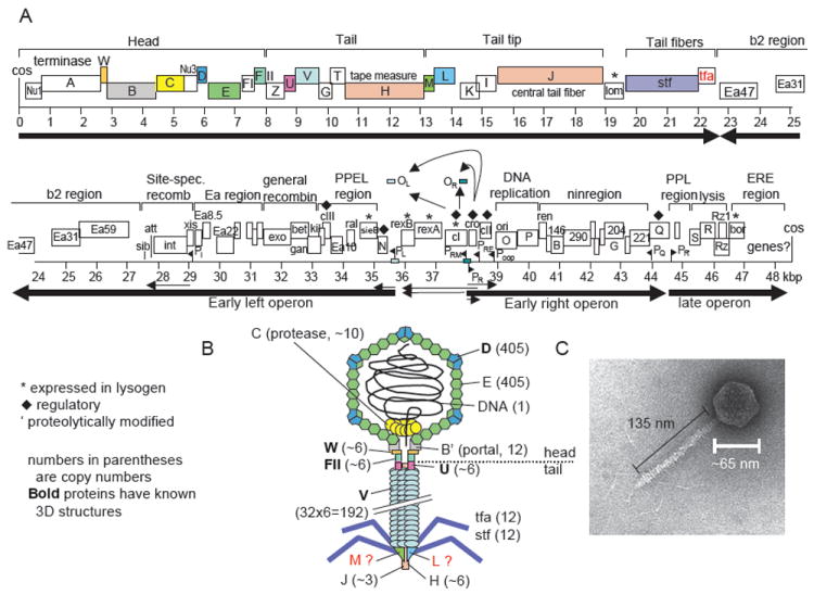
FIGURE 3.

Genome and virion of phage λ. (A) Genome of phage λ. Colored ORFs correspond to colored proteins in (B). Main transcripts are shown as arrows. After Hendrix and Casjens in Calendar (2006). (B) Schematic model of λ virion. Numbers indicate the number of protein copies in the particle. It is unclear whether gpM and gpL proteins are in the final particle or only required for assembly. (C) Electron micrograph of phage λ. Modified after Hendrix and Casjens (2006).
