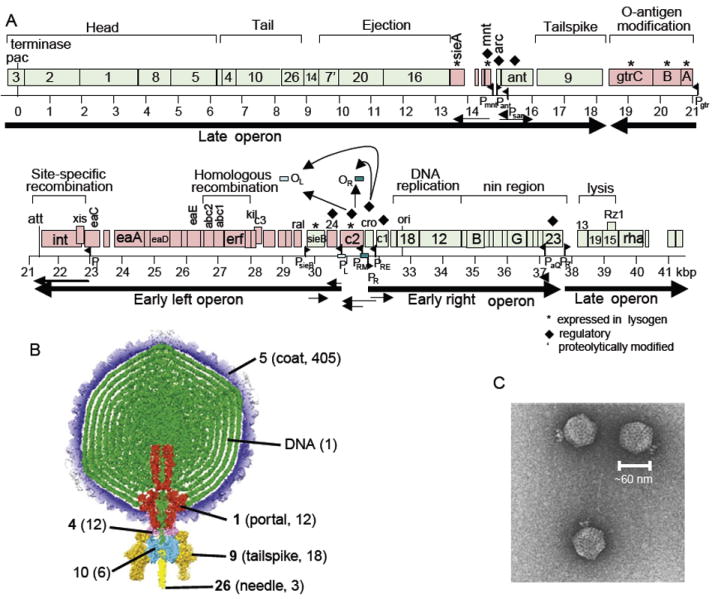
FIGURE 7.

Genome and virion of phage P22. (A) Genome of phage P22 with a scale in kbp below. Green open reading frames (ORFs; rectangles) are transcribed left to right; red ORFs are transcribed right to left. Known RNA polymerase promoters and transcripts are shown as black flags and arrows, respectively. (B) The asymmetric (not icosahedrally averaged) P22 virion three-dimensional cryo-EM reconstruction from Tang et al. (2011). Numbers in parentheses indicate molecules/virion, and bold numbers indicate proteins for which X-ray structures are known. (C). Negatively stained electron micrograph of P22 virions.
