
FIGURE 8.
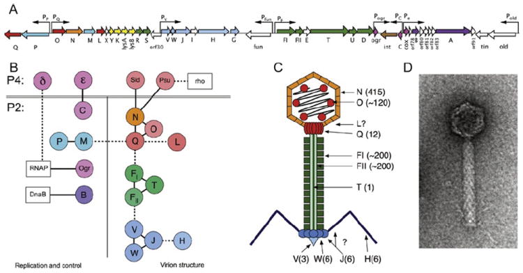
(A) Schematic diagram of the 33.6-kb P2 genome. Open reading frames (ORFs) are indicated by arrows that reflect their direction and size and are color coded as follow: red/orange, capsid-associated proteins (gpQ, O, N, L); blue, terminase proteins (gpP, M); yellow, lysis proteins (gpY, K, lysA, lysB); green, tail-related proteins (gpR, S, FI, FII, T, U, D); blue, base plate and tail fiber-related proteins (gpV, W, J, H, G); pink, transcriptional control proteins (Ogr, C, Cox); brown, integrase (Int); and purple, replication-related proteins (gpA,B). Nonessential genes and ORFs of unknown function are white. Promoters are indicated by arrows above ORFs. (B) Diagram of protein–protein interactions listed in Table IX. Each P2 and P4 protein is shown as a circle, colored as given earlier. Host proteins are shown as white boxes. (C) Schematic diagram of the P2 virion, color coded as given earlier. Copy numbers of proteins, when known, are indicated in parentheses. (D) Electron micrograph of a P2 virion, stained negatively with uranyl acetate.