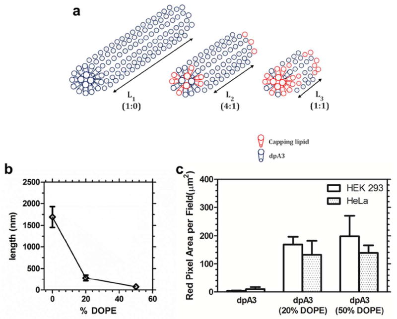
Figure 10. Proposed mechanism for fiber-length by capping phospholipid.

(a) Unsaturated lipid chains of DOPE promote intra-fiber phase separation to the termini of ELPA nanofibers assembled by saturated lipids. (b) The average nanofiber length was plotted as a function of the % of DOPE capping lipid. (c) Plain dpA3 fibers have low propensity for cellular uptake. ANOVA analysis shows a significant difference between the 3 groups (P=0.0188). Tukey’s post hoc analysis shows a significant difference between dpA3 and formulation with 20% & 50% DOPE (P<0.05). No significant difference in uptake between 20% & 50% DOPE was observed. Hence, addition of DOPE increases uptake irrespective of the cell line used. Values indicate the Mean ± SD (n=3)
