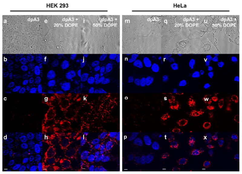
Figure 8. DOPE addition promotes cellular uptake of ELPAs.

16.7μM of particles were added to HEK293 cells and incubated for 5hrs. DiI was used to label nanoparticles (red). DAPI (Blue) was used to stain the nucleus. All images were obtained with the same microscope settings. Image panels (a) to (d) and (m) to (p) show minimal uptake of dpA3 in both cell lines respectively. Conversely, image panels (e) to (l) and (q) to (x) show uptake of dpA3:DOPE particles which suggests DOPE influence in cellular uptake. Control DOPC liposomes did not show any cellular uptake (Fig. S5). Scale bars represent 5 μm.
