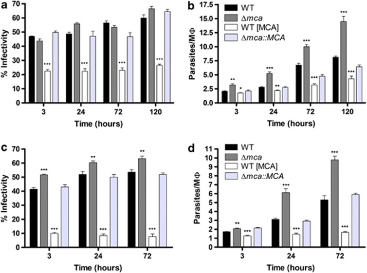
Figure 4.
In vitro macrophage infectivity assay with amastigotes. PEMs were infected with L. mexicana amastigotes from lesions (a, b) or axenic amastigotes, (c, d) at 2 : 1 or 1 : 1 ratio, respectively, for 3 h. The percentage of infected PEMs (a and c) and the number of amastigotes per PEMs (b and d) were determined after 3, 24, 72 and 120 h of incubation. Data show the percentage of infected macrophages and the number of parasites per macrophage (means±s.d from four points) of a representative experiment out of three independent infections. Significant differences compared with WT are indicated as follows (t-test, ***P<0.001; **P<0.01; and *P<0.05)

