
Figure 6.
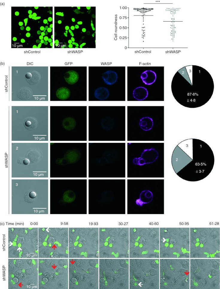
Wiskott–Aldrich syndrome protein (WASP) -deficient CD4+ T cells elongate on intercellular adhesion molecule 1 (ICAM-1) and immature dendritic cells (iDC). (a) Representative images of green fluorescent protein-positive (GFP+) short hairpin (sh) Control and shWASP CD4+ T cells adhering to ICAM-1. Roundness coefficient of 83 shControl and 83 shWASP CD4+ T cells upon adhesion to ICAM-1. Data are from two independent experiments performed with two donors. ***P < 0·001 unpaired t-test. (b) Representative images of GFP+ shControl and shWASP CD4+ T cells in conjugation with ICAM-1-coated beads and immunostained for WASP and F-actin. Percentage of T cells displaying three distinct morphological types defined as follows are represented: type 1: spherical cells with polymerized actin surrounding the bead; type 2: elongated cells; type 3: protruded actin-rich structures away from the bead contact site. Data represent mean ± SEM of two experiments performed with three donors. (c) Movie sequences of shControl and shWASP T cells at the contact of iDC; white arrows indicate cells that round up and red arrows indicate adherent uropods. Results are representative of four experiments performed.