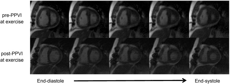
Figure 1.
Representative example of radial k − t real-time images in a patient with predominantly pulmonary stenosis acquired at the same exercise intensity pre-percutaneous pulmonary valve implantation (top row) and post-percutaneous pulmonary valve implantation (bottom row) showing five reconstructed phases including end-diastolic volume (far left) and end-systolic volume (far right). Post-percutaneous pulmonary valve implantation, the left ventricle appears to be better filled (higher end-diastolic volume), with improved right ventricular function. Note the visually better filled left ventricle (increased end-diastolic volume) and reduced right ventricular size.

