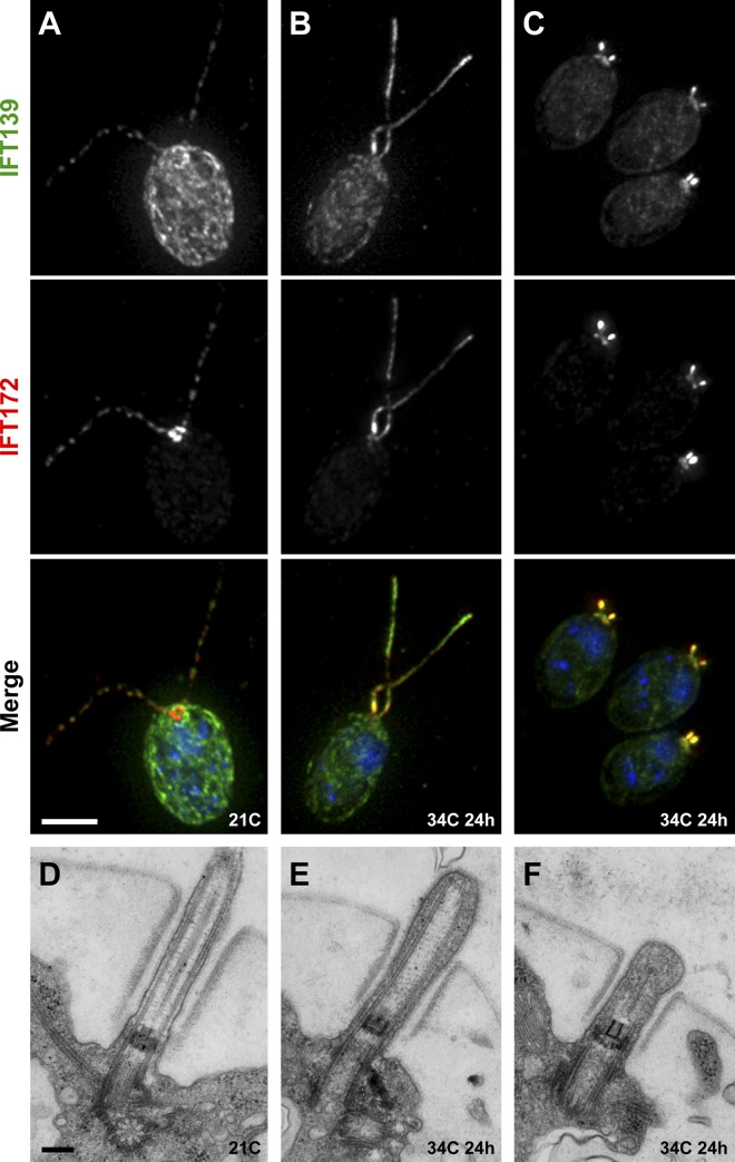
Figure 1.
Temperature-sensitive “retrograde IFT” phenotype of isolated mutant strain dhc1b-3. (A–C) Immunofluorescence and (D–F) transmission electron microscopy (TEM) of the ts flagellar assembly mutant. Green, IFT139; red, IFT172; blue, DNA. At 21°C, the mutant appeared to have a regular distribution of IFT proteins along the flagella (A) and showed no discernible defects in axoneme or basal body ultrastructure (D). After 24 h at 34°C, flagella were either long (B) or stumpy (C) and in both cases showed a strong accumulation of IFT proteins. This accumulation of IFT proteins at 34°C was also apparent by TEM (E and F) as a buildup of electron-dense material between the axoneme and the flagellar membrane. Bars: (A–C) 5 µm; (D–F) 200 nm.

