Figure 5.
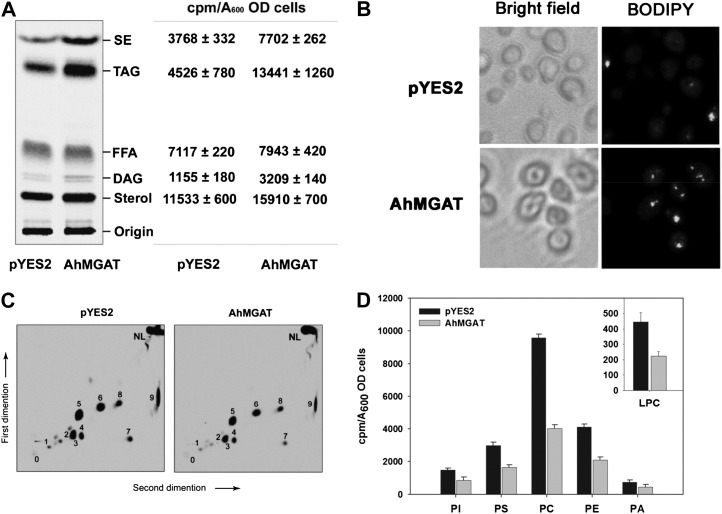
Overexpression of AhMGAT increases TAG formation. MGAT was overexpressed in wild-type yeast in the presence of [14C]acetate (0.5 μCi mL−1). The yeast cells were grown in SM-U containing 2% Gal for 24 h, and equal absorbance (A600 = 20) of cells was harvested and lipids extracted using chloroform:methanol (1:2, v/v). The neutral lipids and phospholipids were separated by silica-TLC, and the radioactivity was quantified by a liquid scintillation counter. A, Phosphor image of TLC showing the [14C]acetate labeling of yeast neutral lipids. Lipids were identified by comparison with known standards. The table represents the incorporation of radiolabels as cpm quantified by liquid scintillation counter using toluene-based scintillation fluid. sd is for three independent experiments. FFA, Free fatty acid. B, Lipid droplet formation in wild-type cells was confirmed by BODIPY493/503 (green) staining. Top panels, wild-type yeast cells expressing pYES2 vector alone; bottom panels, yeast cells overexpressing AhMGAT. C, Incorporation of [14C]acetate into yeast phospholipids, separated by two-dimensional TLC using chloroform:methanol:ammonia (65:25:5, v/v) in the first-dimension solvent system followed by chloroform:methanol:acetone:acetic acid:water (50:10:20:15:5, v/v) as the second-dimension solvent systems. 0, Origin; 1, LPC; 2, PI, phosphatidylinositol; 3, PS, phosphatidylserine; 5, PC, phosphatidylcholine; 6, PE, phosphatidylethanolamine; 4, 7, 8, and 9, unknown; NL, neutral lipid. D, Quantification of phospholipids. Values are means ± sd of three independent experiments. OD, Optical density.

