Figure 9.
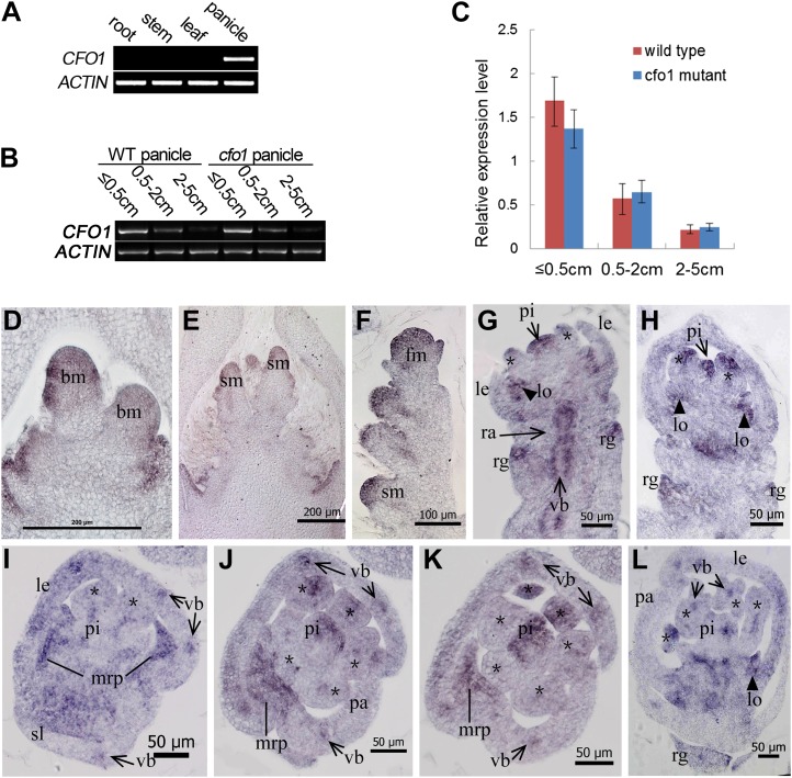
Expression pattern of CFO1. A, CFO1 expression in different tissues as shown by RT-PCR. B, RT-PCR analysis of CFO1 in developing wild-type (WT) and cfo1 panicles at different stages. C, qRT-PCR analysis of CFO1 in developing wild-type and cfo1 panicles at different stages. D to L, In situ hybridization in wild-type panicles and flowers using a CFO1 antisense probe. Transcription of CFO1 was detected in the inflorescence meristem (D and E), spikelet (F), and florets (G–L) at an early development stage. Serial longitudinal sections of one floret are shown in G and H, and serial transverse sections of a different floret are shown in I to K. Asterisks in G to L indicate stamens. bm, Branch meristem; fm, flower meristem; le, lemma; lo, lodicule; pa, palea; pi, pistil; rg, rudimentary glume; sm, spikelet meristem; st, stamen; vb, vascular bundle. Bars = 200 μm in D and E, 100 μm in F, and 50 μm in G to L.

