Figure 3.
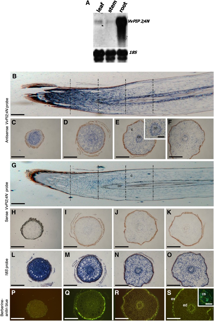
VvPIP2:4N gene expression analyses. A, Northern-blot analysis of VvPIP2;4N aquaporin expression in leaf, stem, and root tissues of cv Nebbiolo. Total RNA was probed with a specific DNA probe corresponding to the full-length VvPIP2;4N gene labeled with DIG. The blots were stripped and reprobed with 18S ribosomal DNA DIG-labeled probe. B to Q, Localization of VvPIP2;4N expression in grape roots. In situ hybridization was performed on longitudinal sections of cv Nebbiolo roots with full-length VvPIP2;4N DIG-labeled antisense (B) and sense (G) RNA probes and on a series of transverse sections (at several distances from the root apex, as indicated by the vertical lines in B and G) with antisense (C–F and E, inset) or sense (H–M) probes. Specific blue signal is mostly evident in meristematic regions and in the regions of vascular differentiation using antisense VvPIP2;4N probe. The sense VvPIP2;4N probe indicates the background level of nonspecific binding in these experiments. In N to Q, a strong signal is evident in the positive controls hybridized with ribosomal Vv18S-DIG-labeled antisense RNA probe. R to U, Fluorescence microscopy of transverse root sections stained with berberine-aniline blue to mark cell wall suberification. The inset in U shows that Casparian bands are evident in the older root region. Bars = 320 μm except in the insets (80 μm). c, Cortical cells; cc, central cylinder; cs, Casparian bands; ed, endodermis; ex, exodermis.

