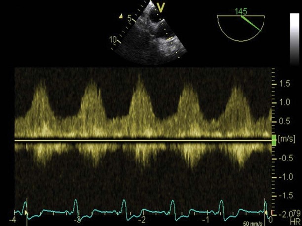
Fig. 4 Transesophageal echocardiogram (pulsed Doppler) focuses on the anastomosis of the left ventricular assist device's outlet cannula to the ascending aorta. Note the typical continuous but highly phasic flow pattern of lower peak systolic velocity (1.5 m/s) in the conduit, which implies the absence of a fixed internal obstruction.
