Figure 1.

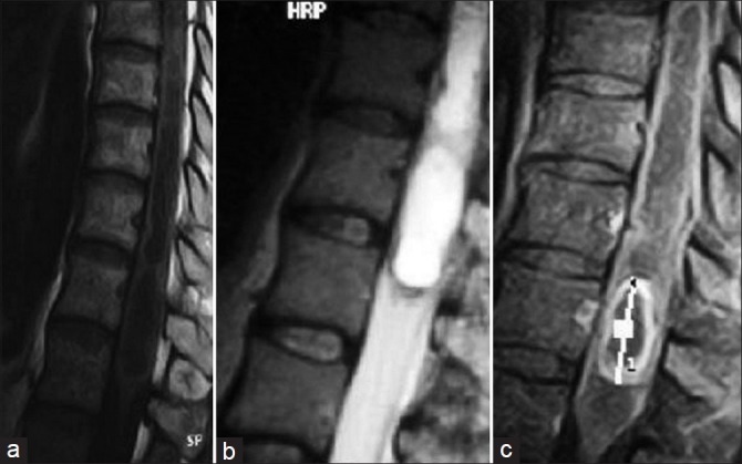
Conus medullaris tumor, which is hypointense on T1‑weighted image and hyperintense on T2‑weighted image, shows peripheral contrast enhancement

Conus medullaris tumor, which is hypointense on T1‑weighted image and hyperintense on T2‑weighted image, shows peripheral contrast enhancement