
Figure 3.
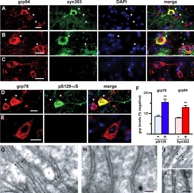
Induction of ER chaperones occurs in neurons with αS pathology. A–E, Confocal immunofluorescence colocalization of grp78 or grp94 with abnormal αS. SpC (A, D), BrSt (B, C), and Ctx (E) sections from end-stage A53TαS Tg mice (A, B, D, E) and nTg (C) mice were double-immunostained for grp94/syn303 (A–C) or for grp78/grp94 (KDEL)/pS129-αS (D, E). Nuclei were stained with DAPI. Neurons showing abnormal accumulation of syn303 or anti-pS129-αS are also associated with increased immunoreactivity for grp78/grp94 (asterisks). Neighboring neurons lacking abnormal αS show lower basal grp78/grp94 immunoreactivity (arrowheads). F, Quantitative analysis obtained using ImageJ software (National Institutes of Health) of neurons with αS abnormalities (stained for syn303 or pS129-αS) in BrSt and SpC of end-stage A53TαS Tg mice shows increased ER stress chaperones (grp78 for pS129-αS, grp94 for syn303). Values are mean ± SEM; n = 13 each for pS129-αS- and syn303-positive neurons and n = 25–31 for negative neurons. **p < 0.001, Student's t test. G–H, Immunogold labeling of αS aggregates shows αS aggregate association with the ER membrane in the diseased A53TαS mice. SpC sections from end-stage A53T (G, I) and nTg (H, J) were immunostained for pS129-αS and processed for electron microscopy. In neurons the pS129-αS-associated gold particles were associated with the ER membranes (arrowheads). No labeling was seen in nTg samples (H, J). The ER cisternae in diseased A53TαS Tg mice were often severely dilated (asterisks) while the ER in nTg controls showed a normal morphology. Scale bar, 50 nm.