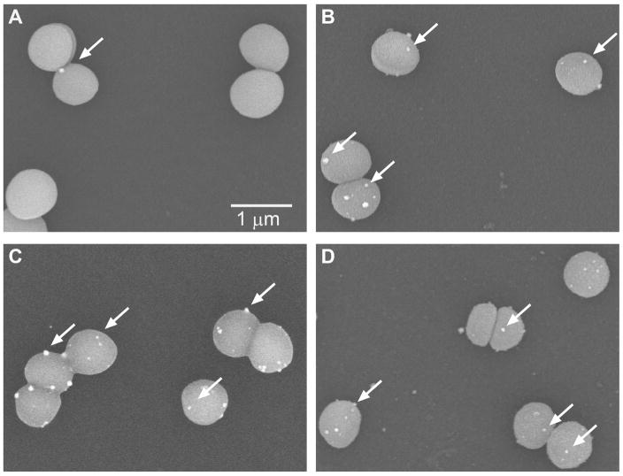
Figure 3.
Scanning electron microscope images of peptide blocking studies. Bacteria were exposed to A)β6-20 peptide, B) no peptide (control), C) β6-20-R14A peptide, or D)β6-20-G15A peptide prior to the addition of the Nanogold-labeled β6-20-NG peptide. Nanogold labels (some are identified by white arrows) are bound to S. epidermidis in the control,β6-20-R14A, and β6-20-G15A blocking samples (B,C,D) while there are minimal labels bound to the bacteria in the β6-20 blocking sample (A).

