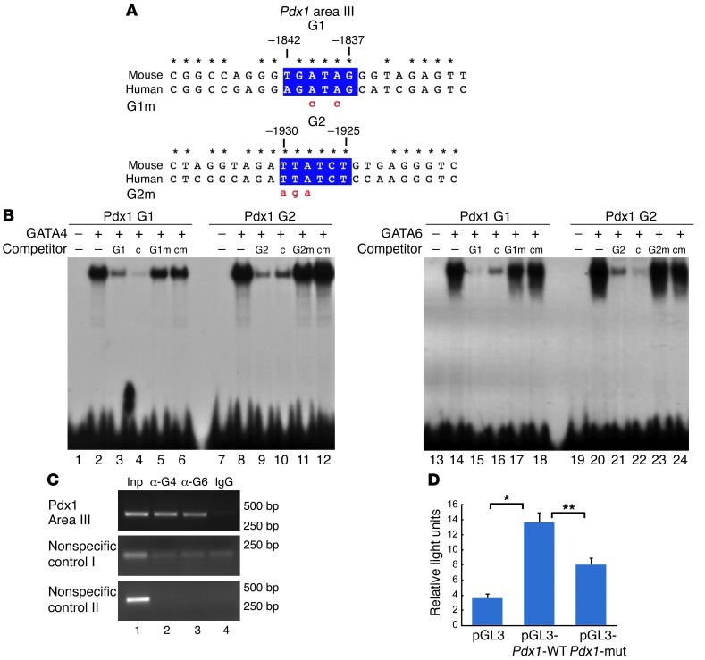
Figure 6. GATA4 and GATA6 bind to the Pdx1 conserved area III in vitro and in pancreatic cell line.
(A) Highly conserved region in the cis-regulatory area III of Pdx1. Two conserved GATA sites, as revealed by bioinformatics analysis, are shown in blue boxes. Numbers indicate the position of the GATA sites relative to the Pdx1 translational start site. Point mutations introduced into GATA sites, G1m and G2m, are indicated in red lowercase. Asterisks denote nucleotides that have been perfectly conserved between mouse and human. (B) Recombinant GATA4 and GATA6 proteins are able to bind to G1 and G2 GATA sites of the Pdx1 enhancer as shown by EMSA. Competition experiments were performed by adding excess unlabeled probes of G1, G2, or control (denoted as c in competitor row) GATA sites, and the corresponding mutant versions (G1m, G2m, or cm) to the binding reaction. (C) ChIP experiments performed in mouse pancreatic ductal cells (mPAC cells) using specific GATA4 and GATA6 antibodies (lanes 2, 3, respectively) and nonspecific anti-IgG (lane 4) show that anti-GATA4 and anti-GATA6 antibodies are able to immunoprecipitate the GATA sites of the Pdx1 conserved area III, but not nonspecific genomic regions. Lane 1 contains PCR products from input DNA (Inp) amplified prior to immunoprecipitation. Sizes of the PCR products in bp are shown on the right. (D) A WT Pdx1 promoter-luciferase construct (pGL3-Pdx1-WT) is significantly activated by endogenous factors present in mPAC cells compared with the activity of the empty reporter pGL3 vector. Mutations in the GATA sites of Pdx1 (pGL3-Pdx1-mut) significantly attenuate the luciferase activity. *P = 0.002; **P = 0.005.

