
Fig. 3.
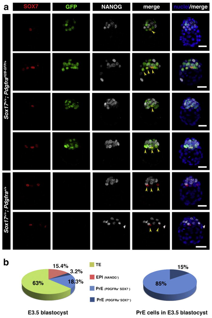
SOX7 is selectively localized to the PrE cells positioned adjacent to the blastocyst cavity prior to lineage segregation. (a) Immunodetection of SOX7 in preimplantation mouse embryos at E3.5 (65–80 cells). SOX7 is first detected in E3.5 embryos after 64-cell stage, localizing only to cells in contact with the blastocoel cavity (arrowheads). In PdgfraH2B–GFP/+ embryos it is co-localized with GFP in cells adjacent to the cavity. Each row represents one embryo, all panels show 3D-reconstruction of confocal images. Pdgfrα-GFP, green; SOX7, red; NANOG, white; Hoechst, blue. Scale bar: 20 μm. (b) Distribution of TE (green), EPI (red) and PrE (SOX7-negative — light blue, and SOX7-positive — dark blue) cells in embryos at E3.5.