
Fig. 5.
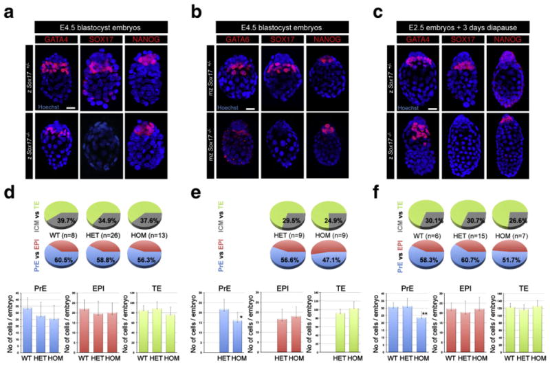
Mouse blastocysts lacking Sox17 correctly specify and segregate embryonic and extraembryonic lineages. Zygotically zSox17 +/− (upper panels) and zSox17−/− embryos (lower panels) at E4.5 (a) and 3 days after tamoxifen injection (c). Maternally and zygotically ablated mzSox17 +/− (upper panels) and mzSox17−/− embryos (lower panels) at E4.5 (b), Blue, nuclei; red, GATA4, GATA6, SOX17 and NANOG. Scale bar: 20 μm. (d–f) Distribution of PrE, EPI and TE cells in Sox17+/+, Sox17+/−, Sox17−/− embryos at E4.5 (d), 3 days after tamoxifen injection (f) and in maternal/zygotic mutant embryos at E4.5 and (e). Blue, PrE; red, EPI; green, TE; grey, ICM. Statistical T-tests are indicated when significant (*:p<0.02; **:p<0.003). Error bars indicate s.d.