
Fig. 7.
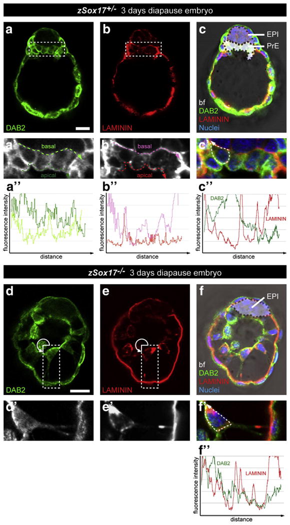
Disrupted organization of primitive endoderm correlates with basement membrane deposition in the absence of Sox17. Immunodetection of DAB2 and Laminin in Sox17+/− (a–c, a′–c′, a″–c″) and Sox17−/− (d–f, d′–f′, f″) 3 day diapause embryos. (a′–f′) magnified views of (a–f) respectively. (a″,b″) Measurements of the fluorescence intensity of DAB2 (a″) and Laminin (b″) staining along the apical (green and red dashed lines) and basal (light green and pink dashed lines) of the PrE layer. (c″,f″) Measurements of the fluorescence intensity of Laminin (red) and DAB2 (green) throughout the cell membrane (dashed line). All panels show single optical section. DAB2, green; Laminin, red; nuclei, blue; bf, bright field. Scale bar: 20 μm.