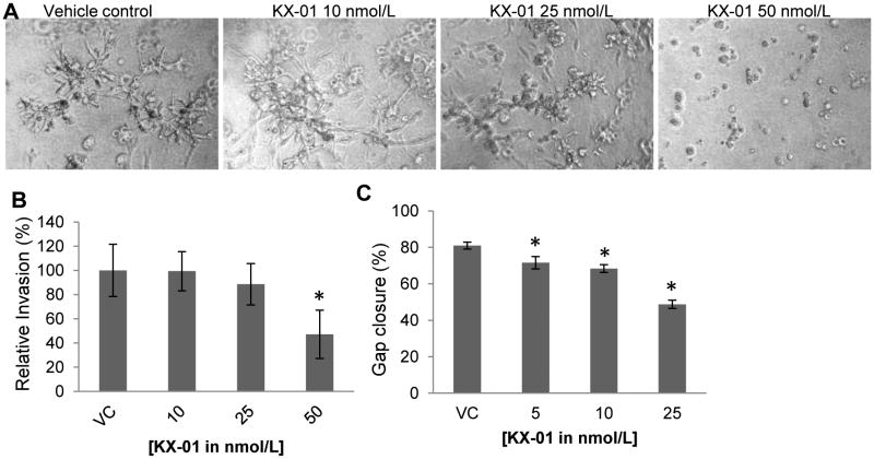
Figure 5.
KX-01 inhibited invasive stellate formation, invasion, and migration of MDA-MB-231 cells in vitro. A) Invasive stellate structures of MDA-MB-231 cells in 3D culture following vehicle or KX-01 treatment were photomicrographed day4. B) MDA-MB-231 cells were incubated with vehicle (VC) or KX-01 (10, 25, 50nmol/L) for 24h and the number of cells invaded was quantified and normalized to cell number and presented as % relative invasion ±SD. *, P<0.05 statistically significant compared to VC. C) Monolayer cultures of MDA-MB-231 cells were gently scratched with a pipette tip to produce a wound. Photographs of cultures were taken immediately after the scratch (0h) and after 24h incubation with vehicle or KX-01 (5, 10, 25nmol/L). Migration is presented as % gap closure ±SD, *, p<0.05, significantly different from VC. Data are representative of three independent experiments.

