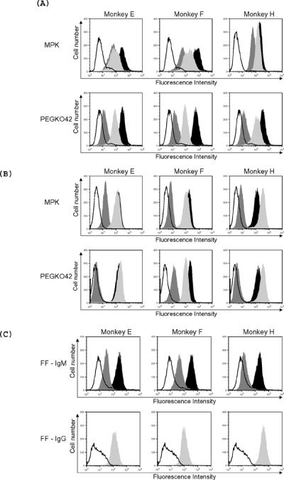
Figure 2. Levels of IgM and IgG xenoantibody binding to wild type and GalTKO pig cells at days 0, 8, and 21 as demonstrated by flow cytometry.

The tracing that is not shaded indicates background binding of secondary antibody alone. Dark gray, black, and light gray filled areas indicate binding at days 0, 8, and 21, respectively. IgM xenoantibody binding to MPK cells (wild type minipig kidney cells) and to PEGKO42 (GalTKO endothelial cells) (A); IgG xenoantibody binding to MPK and to GalTKO endothelial cells (B) and binding of IgM and IgG xenoantibodies to GalTKO fetal fibroblasts (FF, section C) are shown.
