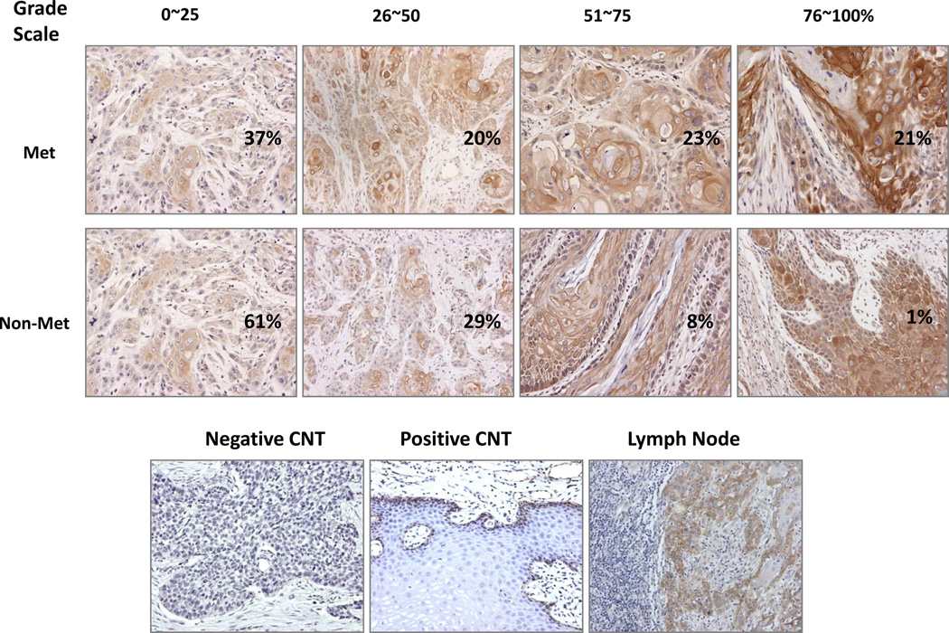
Fig 1.
Integrin β1 expression pattern in HNSCC tissues from patients with and without metastasis. IHC analysis of integrin β1 in HNSCC samples shows membrane and cytoplasmic expression patterns. Of the patients without metastasis, 62% had 0~25% positive staining, 29% had 26~50% positive staining, 8% had 51~75% positive staining, and only 1% of patients had 76~100% positive staining, while of the patients with metastasis, 37% had 0~25% positive staining, 20% had 25~50% positive staining, 23% had 51~75% positive staining, and 21% of patients had 76~100% positive staining. Tissue stained with IgG only and normal epithelium staining were used as the negative and the positive controls, respectively. Metastatic lymph node shows a similar staining as its primary counterpart (Magnification 200 ×).

