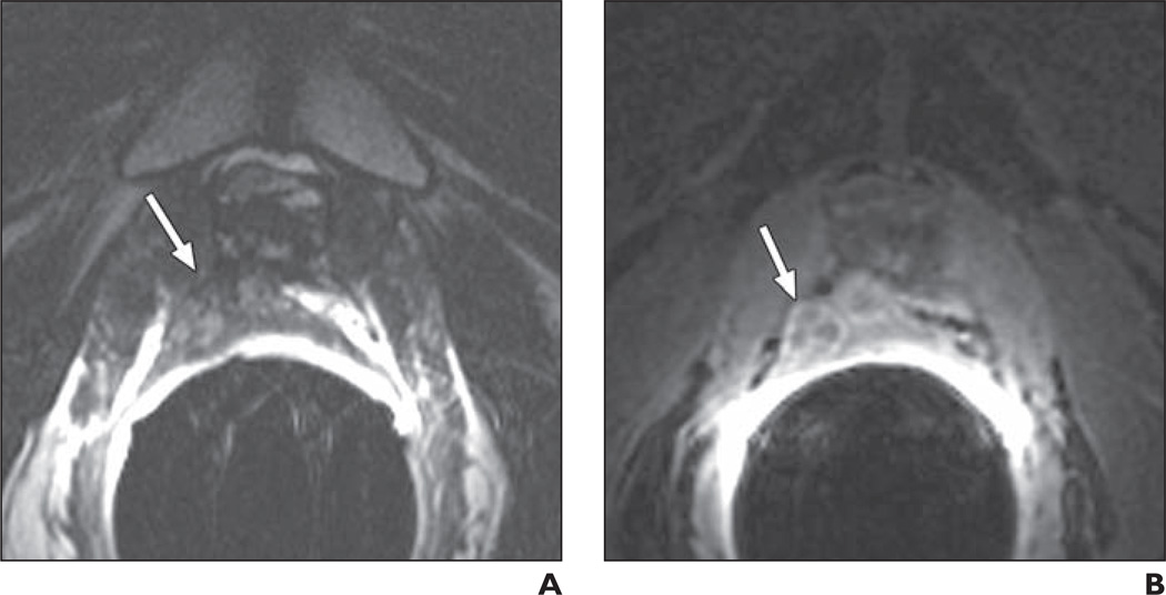
Fig. 5.
66-year-old man 4 years after radical prostatectomy for Gleason 7 (3 + 4) tumor. Follow-up showed biochemical recurrence with elevated prostate-specific antigen of 0.19 ng/mL and palpable nodule at digital rectal examination.
A, Transverse T2-weighted image shows 22-mm nodule (arrow) suggestive of local recurrence in right posterior part of vesicourethral anastomosis.
B, With addition of contrast-enhanced MRI (CE-MRI), level of suspicion did not increase for either of the two readers, both of whom had assigned a score of 5 according to T2-weighted imaging; however, CE-MRI allowed both readers to better demarcate local recurrence (arrow) in relation to surrounding organs. Transrectal ultrasound–guided biopsy verified Gleason score 7 local recurrence.

