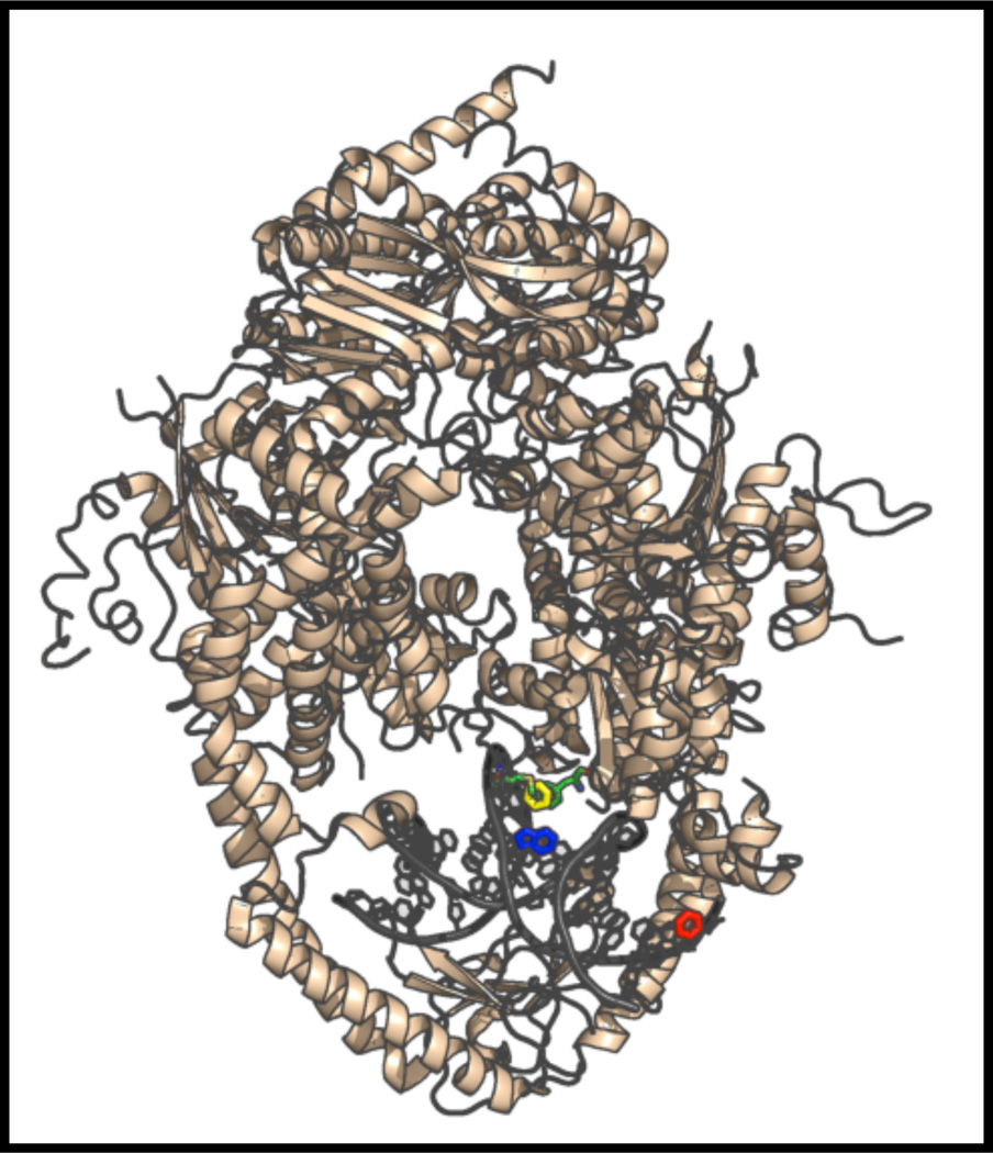
Figure 10.
The X-ray co-crystal structure of Msh2-Msh6 bound to DNA containing a single +Tloop (yellow) with the positions of 6-MI highlighted. The location of 6-MI in the Msh_TFA15 duplex is shown in blue and in the Msh_TFA09 duplex is shown in red. The Phe residue (green) inserts at the +T loop, distorts the DNA backbone and is proximal to the 6-MI in the Msh_TFA15 duplex. Figure was generated with pymol using the coordinates from pdbid, 2O8F.

