
Fig. 3.
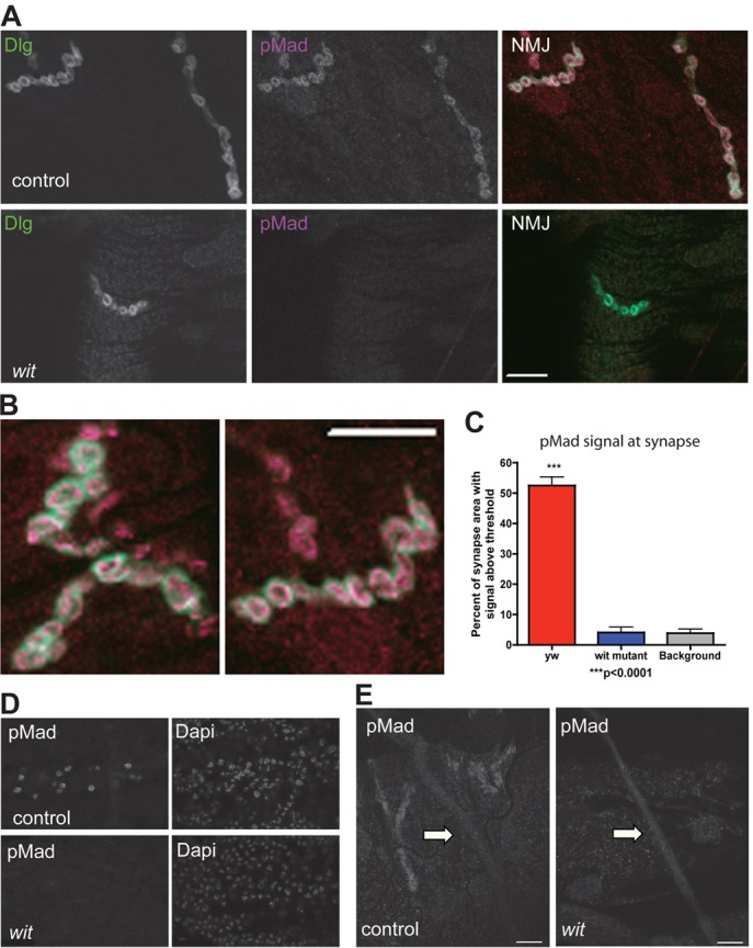
Distribution of phosphorylated Mad in motoneurons. (A) Confocal single section images showing synaptic terminal of muscle 4 in control motoneurons (upper panel, yw) and in motoneurons in the absence of BMP signaling (lower panel, witA12/witB11) stained with antibodies to the post-synaptic protein Discs large (Dlg) and pMad. Note pMad is absent in wit mutants. (B) Confocal single section images at high magnification of control muscle 4 synaptic terminals showing post-synaptic Dlg (green) surrounding the pMad signal (magenta), indicating that pMad is pre-synaptic. (C) Quantification of pMad staining intensity at the NMJ in controls (yw), wit mutants (witA12/witB11) and controls without pMad primary antibody (background staining). (D) wit mutants lose pMad staining in the motoneuron nuclei. Nuclei counterstained with DAPI. (E) No pMad staining above background levels (wit mutant) was seen along the axons (arrows) of control larvae, despite the positive neighboring synaptic staining. Immunofluorescence study of pMad carried out with the PS1-P antiserum (Persson et al., 1998). Scale bars: 10 µm.