
Fig. 4.
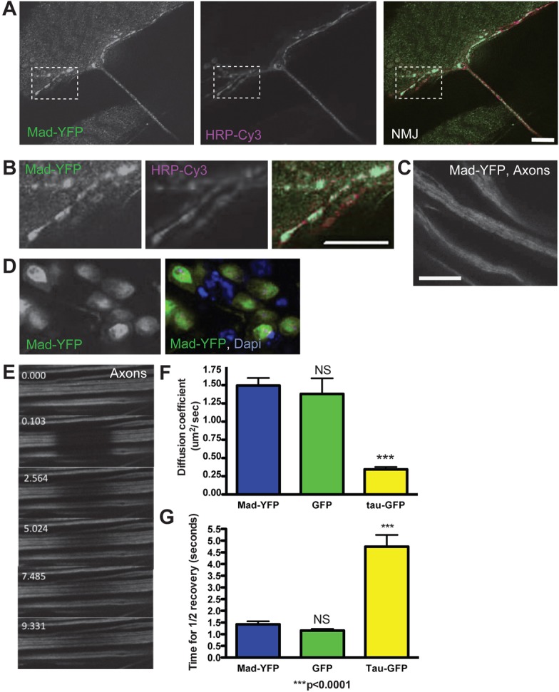
Mad–YFP localizes to the synaptic terminal, axon and cell body. (A) Mad–YFP signal (green) is seen at the synaptic terminal labeled with the presynaptic marker HRP (magenta). (B) An enlarged area of the synaptic terminal (boxed region in A). (C) Mad–YFP is seen along the axon as a diffuse signal. (D) In the motoneuron cell body Mad–YFP localizes predominantly to the nucleus (labeled with DAPI, blue), but it is also present in the cytoplasm. (E) Frames from a time-lapse recording of a FRAP experiment showing axonal Mad–YFP. Recovery of Mad–YFP signal indicates Mad mobility along the axon. The time stamp is in seconds. (F,G) Quantification of FRAP parameters of axonal Mad–YFP shows similar motion parameters to unconjugated GFP and faster movement than the microtubule binding protein Tau–GFP.