
Fig. 6.
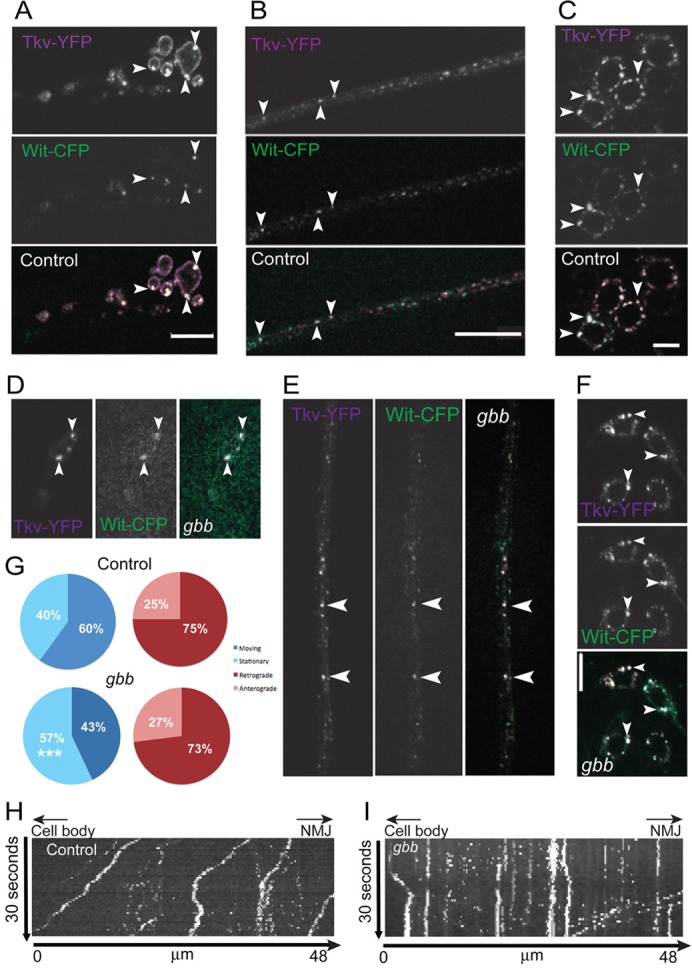
Decreased axonal movement of vesicular receptors in the absence of ligand. (A–C) Coexpressed Wit–CFP (green) and Tkv–YFP (magenta) partially colocalize (arrowheads, white) in motoneuron synaptic terminals (A), motoneuron axons (B) and cell bodies (C) of control animals. (D–F) Colocalization in the NMJ (D), axons (E) and cell bodies (F) is maintained in gbb mutants. (G–I) Quantification (G) of moving and stationary colocalized Tkv–YFP and Wit–CFP vesicles and directionality of axonal traffic in controls and gbb mutants using data from kymographs of controls (H) and gbb mutants (I). Cell body to the left, synaptic terminal to the right. Descending traces represent anterograde traffic, ascending traces retrograde traffic, vertical traces non-motile vesicles. Scale bars: 10 µm.