
Fig. 8.
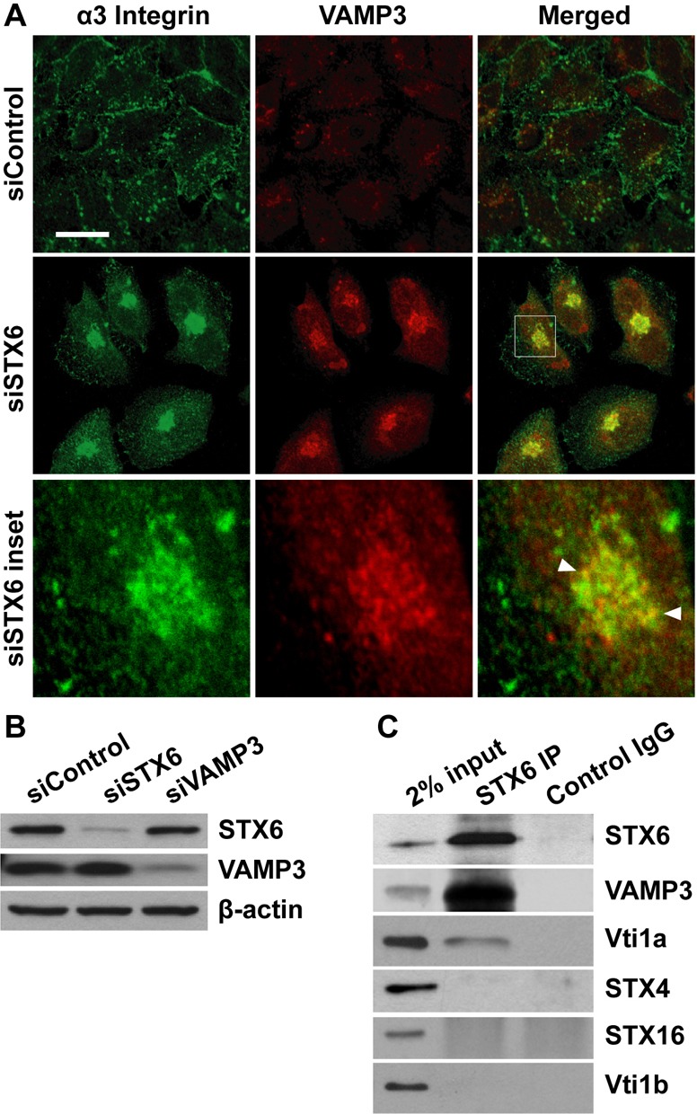
VAMP3 and STX6 form a v-/t-SNARE complex. (A) α3 integrin is accumulated in VAMP3 compartments in STX6 knockdown cells. 60 h after transfection with the control or STX6 siRNAs, HeLa cells were permeabilized and dual stained with an antibody to α3 integrin and an antibody to VAMP3. siSTX6 inset panels: enlarged view of the boxed region in the merged image showing the colocalization (arrowheads) of α3 integrin and VAMP3. Representative single-slice confocal images of three independent experiments are shown. Scale bar: 50 µm. (B) Lysates of HeLa cells transfected with the control, STX6 or VAMP3 siRNAs were immunoblotted with an antibody to STX6, VAMP3 or β-actin. (C) Lysates of HeLa (2 mg) cells were immunoprecipitated with a STX6 antibody or control rabbit IgG, followed by immunoblotting analysis for STX6, VAMP3, Vti1a, STX4, STX16 and Vti1b. 2% (40 µg) input lysates serve as a loading control.