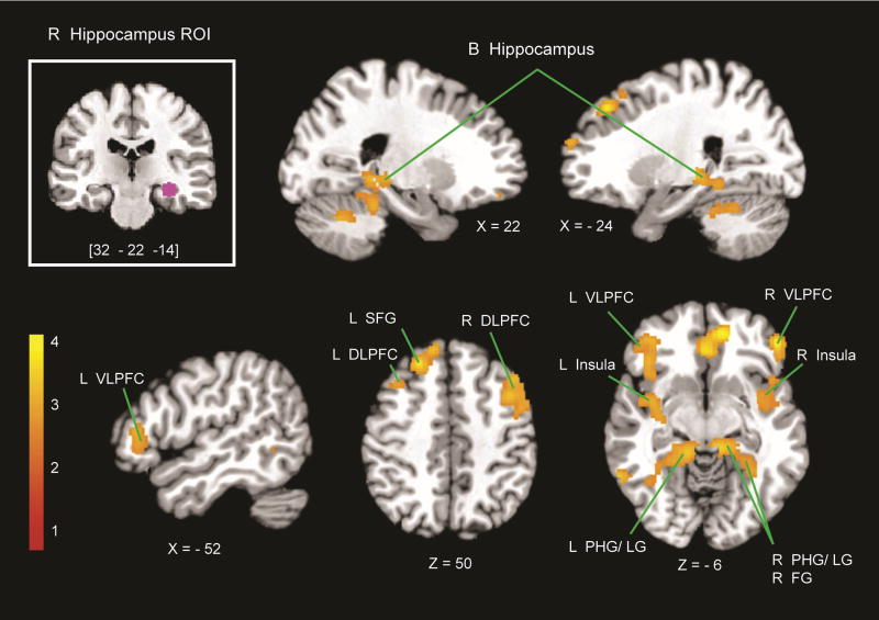
Figure 4.
PPI with the right hippocampus. Using the right hippocampus as an ROI (top-left inset), PPI analyses revealed regions that showed greater effective connectivity with the right hippocampus during the addition, compared with the control task. Greater connectivity was observed in the bilateral hippocampus (B hippocampus), bilateral VLPFC, bilateral DLPFC, left SFG, bilateral insula, bilateral LG, bilateral PHG, and right FG. L = left; R = right.

