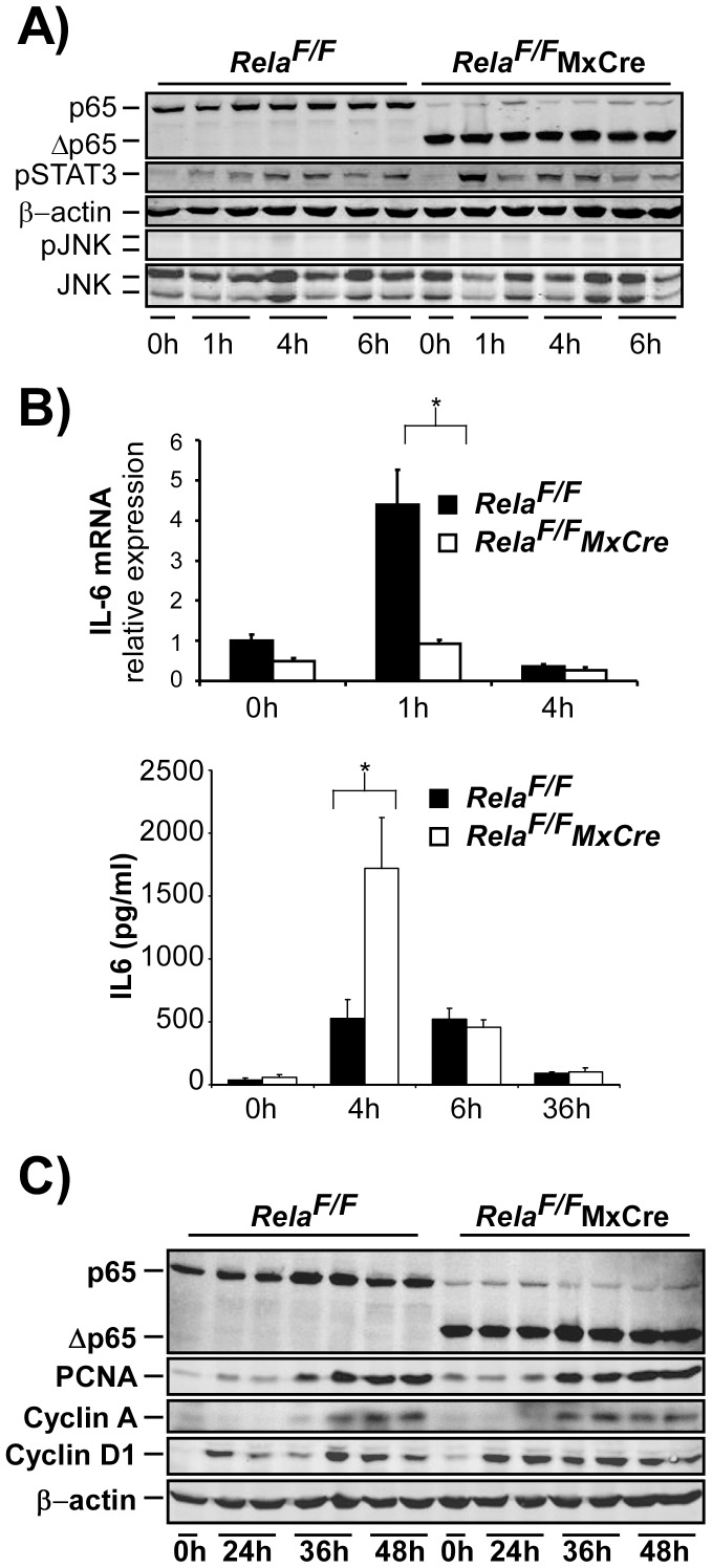
Figure 5. Additional genetic deletion of RelA/p65 in all liver cells in RelaF/FMxCre alters the cytokine response without significantly altering cell cycle progression after 2/3 PH.
(A–C) 2/3 PH was performed on RelaF/F and RelaF/FMxCre animals and analyzed as indicated. (A) Levels of phospho-STAT3 in control and RelaF/FMxCre animals were not different while phosphorylation of JNK was barely detected in either group as assessed be immunoblot analysis. (B) Induction of liver IL-6 mRNA was inhibited in RelaF/FMxCre animals as determined by RT-PCR (upper image), however, IL-6 serum levels were significantly elevated in livers of RelaF/FMxCre mice at 4 h post PH (lower image). (C) Immunoblot analysis of cell cycle associated proteins was performed as described in Fig. 2. Data from cytokine analysis and RT-PCR are presented as the average ± SEM for 3–6 animals per time point per group. *, p≤0.05 for mutant vs. control mice.

