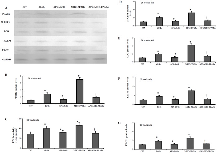
Figure 6. Protein levels of PPARα target genes in hearts of mice at the age of 20 weeks.
Cardiac tissues from db/db mice and MHC-PPARα mice with/without APS administration were detected for the protein expression at the age of 20 weeks, with C57 mice as normal control. Myocardial protein levels of PPARα target genes were determined by Western Blot analysis. (A) Representative autoradiographs of PPARα target genes and GAPDH (loading control) using specific antibodies. Protein levels of PPARα target genes in myocardium, encoding PPARα (B), M-CPF 1 (D), ACO (E), FATP 1(F) and FACS 1(G). PPARα activities in myocardial tissue were also determined at the end of the experiment by RIA (C). All groups n = 8. Bars represented means±S.E.M., and were corrected for GAPDH signal intensity and normalized to the value of C57 wide-type mice. *P<0.05 vs. C57 mice, # P<0.05 vs. untreated db/db mice, † P<0.05 vs. untreated MHC-PPARα mice.

