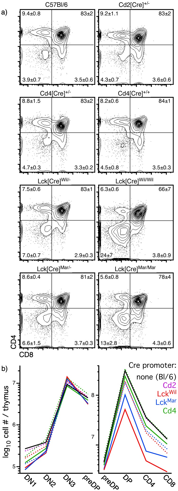
Figure 4. Thymus-initiated Cre expression results in a loss of DP cells.
Panel a) shows the relative proportions of various populations identified by CD4 and CD8 staining on thymocytes from various types of Cre transgenic mice, or C57Bl/6 controls. Statistics indicate mean ± s.d. for 3–9 independent experiments. Panel b) shows absolute numbers of cells per thymus for each major stage of differentiation in the same mice (dashed line = hemizygous; solid line = homozygous); note that because of extensive overlap, only the mean value is shown. Absolute numbers of early progenitor stages were very similar in all strains of mice, suggesting that the effects of Cre were minimal among at these stages. In contrast, substantial differences were seen at the transition to the DP stage. Both strains of hemizygous mice expressing Cre under the Lck promoter showed substantial reductions in DP cell number, changes that are exacerbated in homozygous mice of these strains. These data are in complete agreement with cellularity data shown in Fig. 2, and suggest that the presence of Cre and, in particular, the absolute levels of Cre, may be toxic to DP thymocytes.

