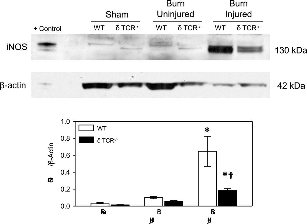
Fig. 1. iNOS expression in uninjured and burn skin lysates.
Skin samples of WT and δ TCR−/− mice at 3 days after burn were assessed for iNOS expression by Western blot as described in Materials and Methods. Raw 264.7 whole cell lysates were used for the positive control (Santa Cruz Biotechnology). Data are mean ± SE for 6–8 mice/group. * p<0.05 vs. uninjured skin or sham skin. † p<0.05 vs. respective WT group.

