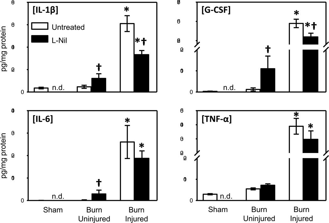
Fig. 4. Effect of iNOS inhibition on skin cytokine content.
Skin samples of untreated and L-Nil treated mice at 3 days after burn were assessed for IL-1β, IL-6, G-CSF and TNF-α expression by Bioplex as described in Materials and Methods. Data are expressed as the mean ± SEM for 7–12 mice/group. * p<0.05 vs. uninjured skin or sham skin. † p<0.05 vs. respective untreated group. n.d. = not determined.

