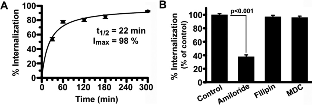
Figure 2. CAM-mediated endocytosis of anti-ICAM nanocarriers by Caco-2 cells.
(A) TNF-α-activated cells were incubated with FITC-labeled anti-ICAM NCs for 30-min to allow binding, non-bound carriers were washed, and cells were incubated for varying times at 37°C to allow internalization. After cell fixation, surface-bound NCs were stained with a TxR-labeled secondary antibody. Fluorescence microcopy was used to analyze the fraction of single-labeled green (internalized) NCs to double-labeled green+red (yellow, surface-bound) NCs. The data were fit by non-linear regression (r2=0.99). (B) Internalization of anti-ICAM NCs (1 h) was assessed in the presence of 3-mM amiloride, 50-µM MDC, or 1-µg/ml filipin, and data was normalized to control. Data are means±S.E.M. (n≥20 cells from 2 experiments).

