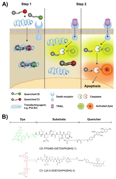
Figure 1.
A) Schematic diagram of imaging the caspase cascade. Conventional dark-quenched, cell-impermeable fluorogenic probes for caspases can be efficiently delivered into the cells followed by simple incubation with a commercially available transfection agent such as PULSin® (Step 1). Once delivered into the cells, highly quenched probes are sequentially activated by expressed target caspases triggered by different initiator pathways, e.g. TRAIL-induced caspases activation (Step 2). B) Chemical structures of fluorogenic probes targeting caspase-8 and caspase-3, C8 and C3, respectively. Each probe consists of a substrate and a pair of dye/dark quencher with no significant spectral overlap of emission between the two probes.

