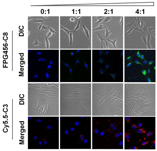
Figure 4.
Fluorescence images of MDA-MB-435 cells incubated with non-quenched FPG456-C8 or Cy5.5-C3 formulated with various amounts of PULSin (1 μg/100 μL of peptides with 0, 1, 2 and 4 μL of PULSin from a commercially provided stock, denoted as 0:1 to 4:1). Each formula was incubated with the cells in 1 mL of L-15 medium without FBS for 3 hr at 37 °C. The cellular uptake of FPG456-C8 and Cy5.5-C3 were examined with a fluorescence microscope configured for FITC and Cy5.5 filters followed by DAPI staining at room temperature. Blue (DAPI), nucleus; green (FPG456); red (Cy5.5-C3).

