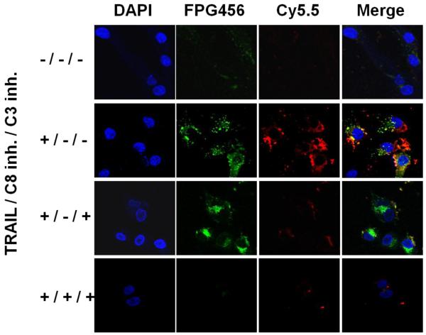
Figure 5.
Multiplexed imaging of the caspase cascade in MDA-MB-435 cells. The cells were incubated with C8/C3/PULSin for 4 h and treated by different combinations of TRAIL (100 ng/mL) and 50 mM of caspase-3 and/or caspase-8 inhibitors. The cells were imaged using confocal microscopy followed by DAPI stating. Blue (DAPI), nucleus; green (FPG456), caspase-8; red (Cy5.5), caspase-3.

