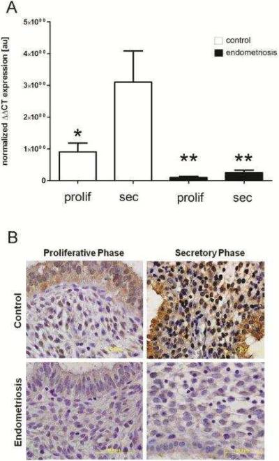
Figure 1. Progesterone-mediated endometrial CB1-R mRNA and protein expression is disrupted in women with endometriosis.
Human endometrial tissues were obtained by biopsy during the proliferative or secretory phase from women with or without endometriosis and a portion flash-frozen for mRNA analysis and a portion formalin-fixed and paraffin embedded for immunohistochemical analysis. Quantitative RT-PCR analysis of CB1-R mRNA expression (A) revealed that in samples obtained from disease-free women, CB1-R mRNA expression was dramatically increased in the secretory phase (N=11) compared with samples obtained during the proliferative phase (N=9). Endometrial tissues obtained from women with endometriosis expressed minimal CB1-R mRNA in both the proliferative (N=7) and secretory (N=8) phases of the cycle. qRT-PCR data, normalized to the housekeeping gene (ribosomal protein large P0, RPLP0), was analyzed using the ΔΔCT method [59] and * is indicative of P<0.05. Immunohistochemical localization of CB1-R (B) was similar to mRNA expression patterns in that protein expression was weak in control samples obtained during the proliferative phase, but robust staining was observed in secretory phase samples. However, CB1-R immunolocalization was minimal in both proliferative and secretory phase samples from women with laparoscopically-confirmed endometriosis. All slides were photographed at 1000× and results representative of at least 5 samples in each group.

