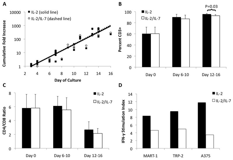
Figure 2.

A. Growth curves for MDLN grown under two culture conditions. MDLN were incubated with anti-CD3/anti-CD28 beads with either IL-2 or IL-2 + IL-7. MDLN growth with either IL-2 or IL-2/IL-7 showed no difference in expansion, achieving over 100-fold expansion by 14 days. R2 ≥ 0.90 for both trendlines. N=10. B. Percent of MDLN cells expressing CD3. MDLN cultures started with 60% CD3+ cells, which increased to 96% CD3+ for IL-2 culture and 93% for IL-2/IL-7 culture (p=0.03 for IL-2 vs. IL-2/IL-7). Error bars indicate 95% CI. N=10. C. Ratio of CD4+ T-cells to CD8+ T-cells. MDLN cultures started with CD4+/CD8+ ratio of 6, which decreased to 3 for IL-2 and 2 for IL-2/IL7 (p=0.43 for IL-2 vs. IL-2/IL-7). Error bars indicate 95% CI. N=10. D. Antigen-specific IFN-γ production. MDLN cells from single patient were incubated alone, with melanoma peptides (MART-1 or TRP-2), or with melanoma cell line (A375) for 72 hours. The stimulation index (SI) for MART-1, TRP-2 and A375 for MDLN cultured with IL-2 was 8.3, 9.5, 11.8, respectively. This was greater than the SI for IL-2/IL-7 (4.6, 5.0, 3.5, respectively). Stimulation index (SI = stimulated sample/MDLN alone sample).
