Figure 3.
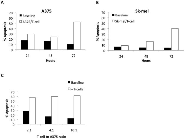
A and B. Determining the optimal time frame for apoptosis assays with MLDN cultures. Day 14 MDLN cultures were incubated with two human melanoma tumor lines (A375 and Sk-mel, respectively) at a MDLN cell to tumor cell ratio of 2:1 for 24, 48 and 72 hours. Tumor cells without MDLN cells were used for baseline apoptosis. Baseline apoptosis was 17.8%, 16.6%, and 10.4% for A375 and 6.8%, 5.2%, and 5.0% for Sk-mel at 24, 48, and 72 hours, respectively. MDLN cells increased apoptosis 11.7%, 7.5%, and 42.4% over baseline for A375 and 2.1%, 11% and 34.9% for Sk-mel at 24, 48 and 72, hours, respectively. C. Apoptosis increases as MDLN cell to tumor cell ratio increases. Day 14 MDLN cultures were incubated with human melanoma cell line (A375) for 72 hours and increasing MDLN cell to tumor cell ratios (2:1, 4:1, 10:1). Apoptosis increased 28.9%, 43.1%, and 48.7% over baseline with increasing MDLN cell ratio.

