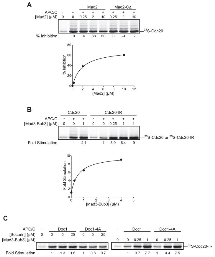
Figure 2. Checkpoint proteins regulate Cdc20 binding to the APC/C.
(A) 35S-Cdc20 in reticulocyte lysate was pre-incubated with the indicated Mad2 or Mad2-CΔ concentration and added to TAP-APC/C beads. Following a 30 min incubation, beads were washed and the bound Cdc20 was analyzed by SDS-PAGE and autoradiography. Results are representative of two independent experiments.
(B) 35S-Cdc20 or 35S-Cdc20-IR in reticulocyte lysate was incubated with the indicated Mad3-Bub3 concentration and TAP-APC/C beads, and analyzed as in panel (A). Similar results were obtained in three independent experiments.
(C) 35S-Cdc20-IR in reticulocyte lysate was incubated with the indicated concentration of securin fragment (aa 1–110) or Mad3-Bub3 and TAP-APC/C beads immunopurified from DOC1 or doc1-4A strains. Results are representative of two independent experiments.

