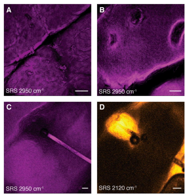
Fig. 5. SRS skin imaging in living humans.

(A-C) SRS images of the stratum corneum and viable epidermis tuned into CH3 stretching vibration of proteins (2950cm-1) showing nuclei of variable size (A and B) as well as a hair (C). (D) SRS image of DMSO penetrating the skin at the same region as (C). We find that DMSO also accumulates in the hair shaft. We used deuterium labeling to create a unique vibration of d6-DMSO at 2120 cm-1 for specific imaging. Images are acquired in epi-direction on the forearm of a volunteer. Image acquisition time is 150 ms for (A) and (B) and 37 ms for (C) and (D), all with 512 × 512 pixel sampling. Scale: 50 μm.
