Figure 4.
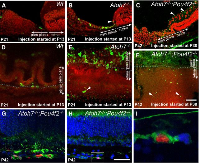
BrdU incorporation and neurogenesis in RGC-depleted retinas. A–C, Cryosections showing the retinal and ciliary margins of representative control retina and RGC-depleted retinas. For A and B, BrdU was injected daily for 9 consecutive days starting at P13 and the retinas were collected at P21. For C, BrdU was injected daily for 12 consecutive days starting at P30 and the retinas were collected at P42. A, In Atoh7lacZ/+ retinas, 9 d of BrdU labels only a single cell at the pars plana. B, In Atoh7lacZ/GFP retinas, 9 d of BrdU from P13–P21 labels numerous cells in the pars plana as well as cells more distally within the ciliary body. One BrdU-labeled cell is visible in the retinal margin (arrowhead). C, BrdU labeling from P30 to P42 labels cells in the ganglion cell layer (arrowheads) and ciliary body. BrdU-positive cells in the pars plana appear more widely spaced. D–F, Flat-mounted retinas showing the retinal and ciliary margins corresponding to the same experimental conditions and genotypes as in A–C. In A–F, Red, Propidium iodide; green, BrdU. G–I, Confocal images of retinas with 99% RGC depletion labeled with BrdU (red) and neurofilament (NF-L, green) from P30 to P42. Nuclei are counterstained with TOPRO-3 (blue). G, Clusters of BrdU-labeled cells in the RGC layer are positive for the neuronal marker NF-L. H, A BrdU-positive cell colabeled with NF-L located in the RGC layer close to the retinal margin. I, Higher magnification of the boxed area in H, shown as a single optical section. Scale bars, 100 μm.

