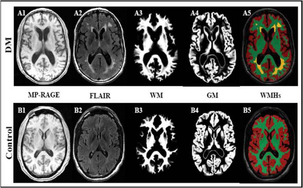
Figure 4.
Anatomical images of a diabetic (A1–A5) and a control subject (B1–B5) demonstrate that type 2 diabetes is associated with atrophy of gray (GM) and white matter (WM), enlarged ventricles) and diffuse periventricular hyperintensities (WMHs). 3D Magnetization prepared rapid gradient echo (MP_RAGE) and fluid attenuated inversion recovery (FLAIR) images were acquired at 3 Tesla MRI (MP_RAGE: TR/TE/TI = 7.8/3.1/600 ms, 3.0 mm slice thickness, 52 slices, bandwidth = 122 Hz per pixel, flip angle = 10°, 24 cm × 24 cm FOV, 256 × 192 matrix size), FLAIR (TR/TE/TI = 11000/161/2250 ms, 5 mm slice thickness, 30 slices, bandwidth = 122 Hz per pixel, flip angle = 90°, 24 cm × 24 cm FOV, 256 × 160 matrix size).

