
Figure 5.
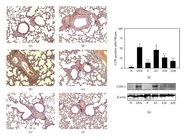
Acacetin reduced COX-2 expression in the lungs. COX-2 expression was analyzed by IHC staining (brown, indicated by arrows) in normal (a), OVA (b), prednisolone (c), A5 (d), A10 (e), and A20 (f) groups (100x magnification). Results were expressed as the number of COX-positive cells per 100 μm (g). COX-2 protein levels were detected by Western blots in lung tissue (h); β-actin expression was used as an internal control. Data are presented as means ± SEM. **P < 0.01 compared with the OVA group.