Figure 1. .
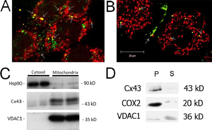
Connexin 43 localizes to mitochondria in RRECs. (A) Immunofluorescence stains of RRECs pretreated with MitoTracker Red and stained with anti-Cx43 antibody showed colocalization of Cx43 on mitochondria. (B) RRECs transfected with Cx43-GFP plasmid show Cx43 localization on mitochondria (stained with MitoTracker Red) in live RRECs. Open arrowheads indicate Cx43 localization on mitochondria. Closed arrowheads indicate Cx43 localization in gap junction plaques between cells. (C) Western blot shows Cx43 localization in protein isolated from mitochondria of RRECs. Hsp90 is used as cytosolic marker, while VDAC1 is mitochondrial marker. (D) Cx43 is predominately present on the inner mitochondrial membrane. COX-2 is used as control for mitochondrial inner membrane fraction, and VDAC1 is used as control for outer mitochondrial membrane fraction. P = pellet representing mitochondrial inner membrane fraction, S = supernatant representing mitochondrial outer membrane fraction, Cyto represents cytosolic fraction.

