Figure 5. .
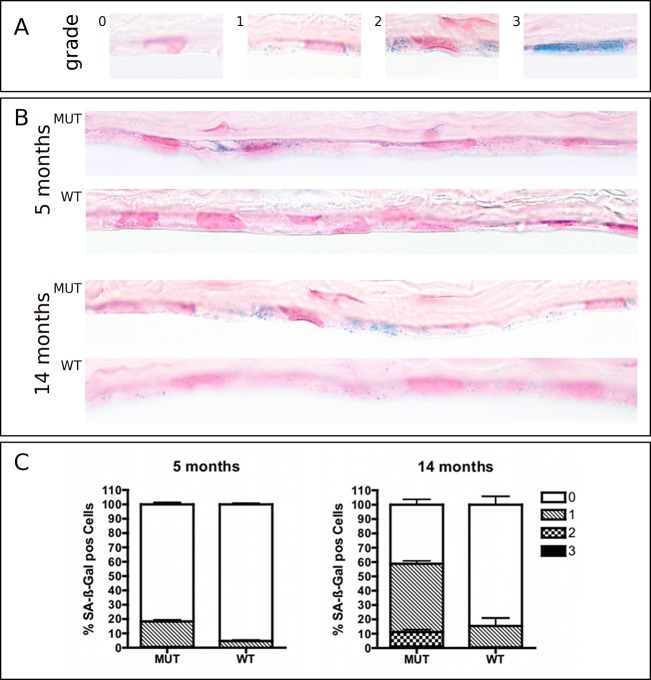
SA-β-Gal staining of corneoscleral buttons from 5- and 14-month-old Col8a2 Q455K/Q455K MUT and WT mice (pH 5.5). Cryosections were counterstained with nuclear fast red. (A) Staining intensity was graded according to a scoring system previously described by Mimura and Joyce14: 0, no staining; 1, focal weak staining; 2, multifocal moderate staining; or 3, multifocal intense staining. (B) Increased staining intensity was noted in sections of MUT compared with WT mice accumulating with age in both strains. (C) Diagrams depict proportions of corneal endothelial cells graded for SA-β-Gal staining in MUT and WT mice at 5 and 14 months. Data are mean ± SEM.

