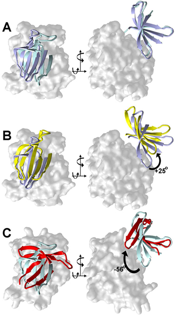
Figure 8.
A comparison of the canonical molecular motions described for the N-lobe core β-sheet in PKA between the open and closed conformations, with the motion in RSK2 on going from the AMP-PNP32 to the SL0101-bound state, described in this paper. The proteins are superposed on the C-lobe, which is shown as solid surface; the core five-stranded β-sheet on the N-lobe is shown as a cartoon.
(A) A comparison between the AMP-PNP-bound structures of PKA (blue) and RSK2 (cyan);
(B) A comparison of PKA structures with AMP-PNP (blue) and in nucleotide-free state (yellow)64;
(C) A comparison of mRSK2NTKD with AMP-PNP (blue) and with SL0101 (red)

